Case Details
Lesion Characteristics
Lesion Size (diameter): 9 mm
Lesion Location: Right Middle Lobe
Bronchus Sign: Yes
Visible on Fluoro: No
REBUS Verification: Concentric Rebus Confirmation
Case Information
Time to First Biopsy: 13 minutes
Full Procedure Time: 30 minutes
ROSE: No ROSE on-site
Definitive Diagnosis: Adenocarcinoma
Background
A 67-year-old woman, non-smoker. CT completed due to unexplained weight loss.
Planning
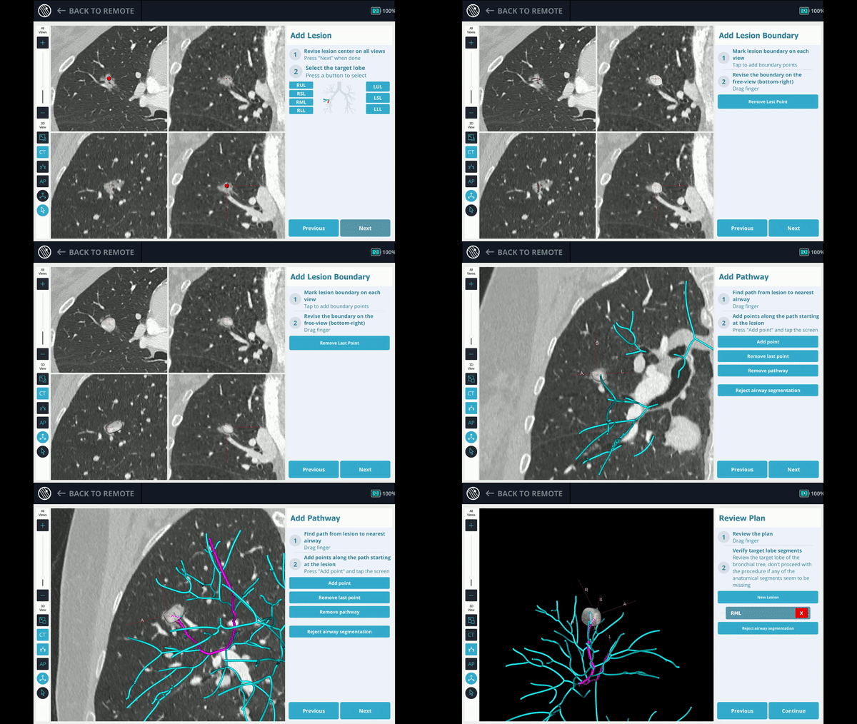
In Planning, the pre-operative CT was uploaded and the lesion and lesion boundaries were marked. The Body Vision System proposed different pathway options to reach the lesion and Dr. Abramovich chose the appropriate one.
Virtual Navigation
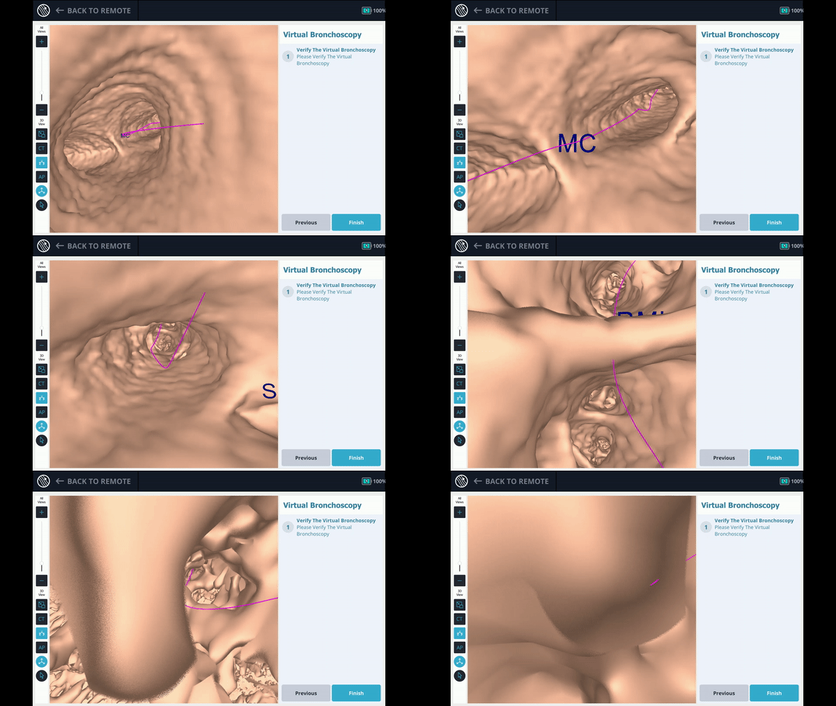
Dr. Abramovich viewed and verified the virtual pathway via Body Vision's virtual bronchoscopy.
CABT Registration
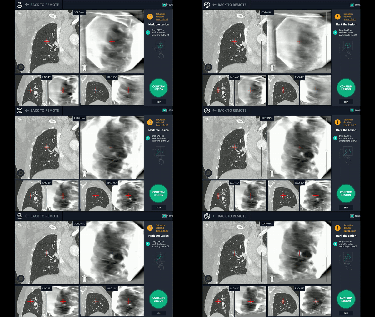
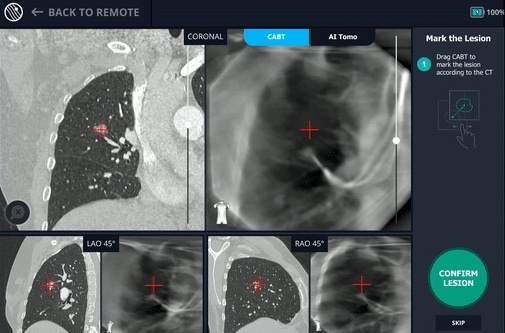
Using only a conventional C-arm, Body Vision produced a tomographic reconstruction of the lesion in 3D, thus enabling Dr. Ambramovich to see the real-time, accurate lesion location and overcome any concerns with CT-to-body divergence.
Tool-in-Lesion CABT
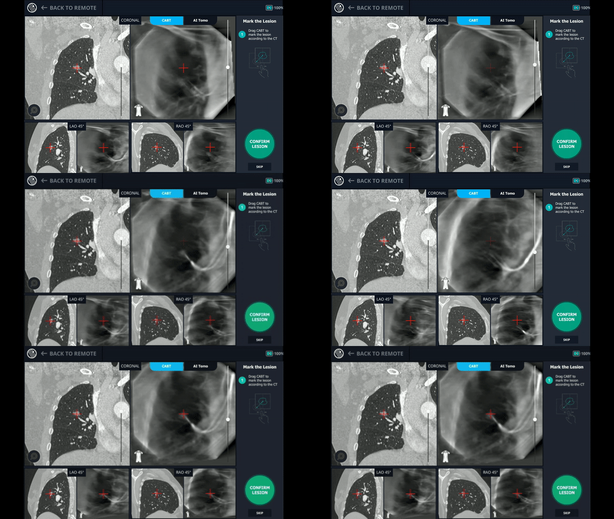
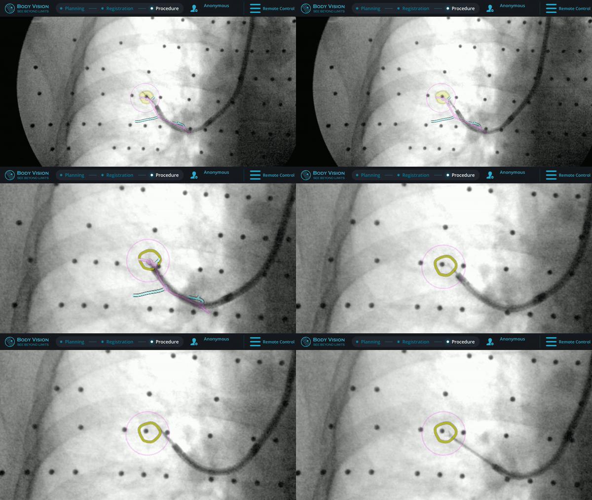
Here, Dr. Abramovich was able to confirm the tool was inside the lesion using Body Vision's real-time, CABT technology.
Biopsy
Dr. Abramovich successfully takes a biopsy sample from within the lesion using forceps.
Conclusion
Small nodules should be diagnosed even if the patient does not have risk factors. The Body Vision system is capable of assisting a definitive diagnosis by creating a reliable augmented fluoroscopy image and providing real-time, CABT tool-in-lesion confirmation in nodules as small as only a few millimeters.
About Dr. Amir Abramovich
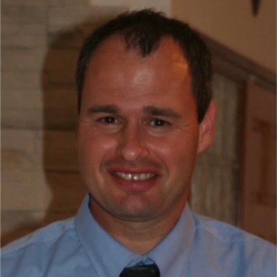
Director, Interventional Pulmonology
Carmel Medical Center
